THERMOGRAPHIC NEUROLOGY
Thermographic Neurology
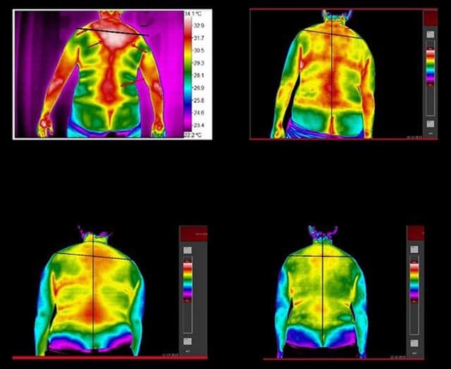
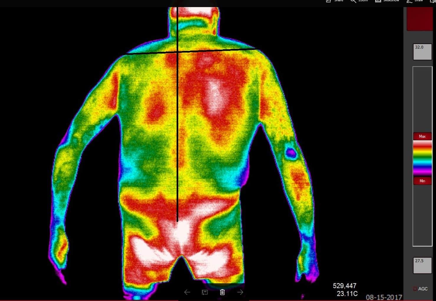
There is a large amount of literature on thermographic neurology. The AMA created an in depth paper in 1987, as this technology was emerging. The paper in itself is a great tool and still the basis of our protocols in thermographic neurology today. The analysis of the hyperthermic and hypothermic patterns allow the Thermologist to make neuromuscular assessments.
Thermography neurology is important in aiding the diagnosis of many diseases that have been difficult to assess with traditional methods. These include conditions such as: radiculitis, peripheral neuritis, complex regional pain syndrome (CRPS), upper motor neuron lesions, multiple sclerosis, ALS, and more neurological diseases.

Thermography has long been used by Neurologists, Chiropractors, and other professional to assess the nervous system. Take a look at the images below. Please read the AMA Council for NMS Thermography
Case Studies
How Thermography Identifies Neurology
The nervous system controls every aspect of the human body, and most pertinent to medical thermology, is the skin surface temperatures. The skin is a complex organ innervated by millions of nerve endings that control capillary function.
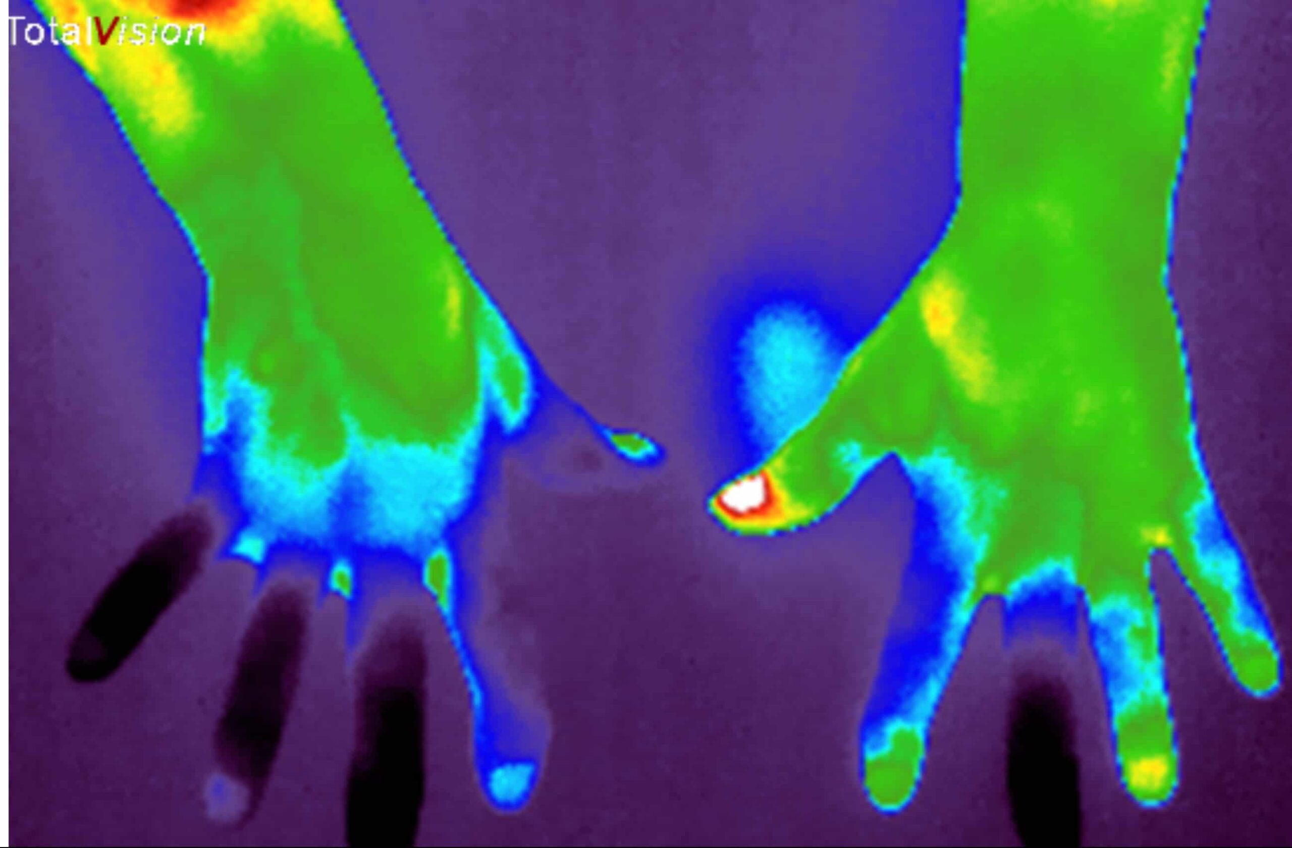
During a sympathetic response the nerves controlling the capillaries with cause vasoconstriction. This in turn will create hypothermia of the tissue innervated. This cold pattern can be plotted on a computer and an assessment can be made.
In the case of Complex Regional Pain Syndrome (CRPS), formerly known as Reflex Sympathetic Disorder (RSD), the sympathetic nervous system is out of control. We test for CRPS will a process called Cold Stressing that looks for inconsistencies with the normal sympathetic response.
A trained Thermologist will assess these neuropathies and put all findings in to a report for the patient.
